Turn your ophthalmic images into clinical answers automatically
From retinal screening to corneal graft assessment, ADCIS builds AI-powered software that transforms raw ophthalmic images into quantitative, reproducible clinical data. Trusted by researchers, clinicians, and device manufacturers in 50+ countries.
Our business? Computer vision : automatically extracting clinical, quantified, and reproducible information from images of the retina, cornea, and surface of the eye!
The numerous projects in which ADCIS has participated have enabled us to develop solutions, partnerships, and expertise covering the entire depth of the eye.
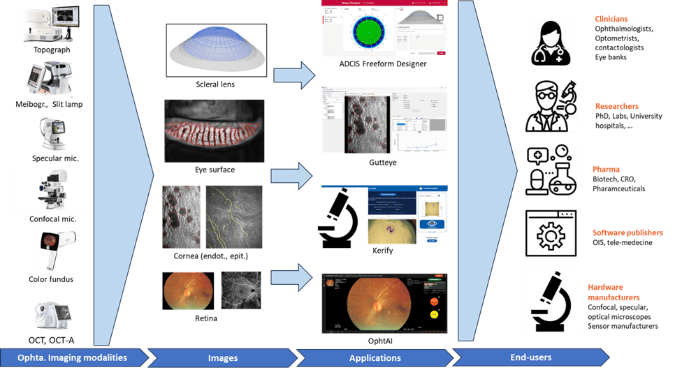
ADCIS solutions in ophthalmology
ADCIS publishes and distributes its own diagnostic software for ophthalmology. Co-developed with its clinical and academic partners, its solutions are available off-the-shelf and target various organizations in the field of ophthalmology:

Eyecare clinical staff
Ophtalmologists, optometrists, contactologists
Get standardized, AI-powered analysis for your clinical routine without changing your imaging setup

Research group
Labs, University, R&D centers
Publication-grade image analysis co-developed with leading academic institutions worldwide

Pharmas
Pharmas, Biotechs, clinical & pre-clinical CROs
Validated, reproducible imaging endpoints for your clinical trials from pre-clinical to Phase III.

Software publisher
Integration of ADCIS modules into business software for healthcare professionals
Integrate AI-powered image analysis into your platform via SDK in weeks, not months

Hardware manufacturer
Integration of ADCIS modules into imaging modalities (fundus, OCT, specular, etc.)
Embed quantitative analysis directly into your device for a smarter, more competitive offering
Les produits ADCIS d'analyse d'images de la rétine

OphtAI
Screening for DR, AMD and glaucoma is time-consuming and specialist-dependent. OphtAI analyzes any fundus image in under a second and flags what needs attention.
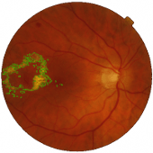
ReV Analyzer
Software for quantifying the volume of pigment epithelial detachments in OCT images of the retina
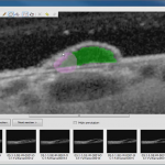
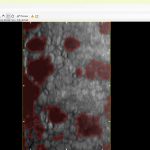
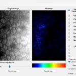
ADCIS corneal image analysis products
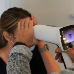
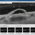
Gutteye
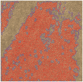
Fuchs dystrophy grading is still done manually, with high variability between operators. GuttEye segments and grades guttae automatically from specular microscopy images in seconds.
CornAI
After a corneal graft, will the cells survive? CornAI predicts graft success at Year+1, +2 and +3 using deep learning on post-operative specular images.
Kerify
Manual estimates of corneal tissue damage are slow and inconsistent. Kerify calculates the area of cell damage automatically so eye banks waste less tissue.
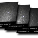
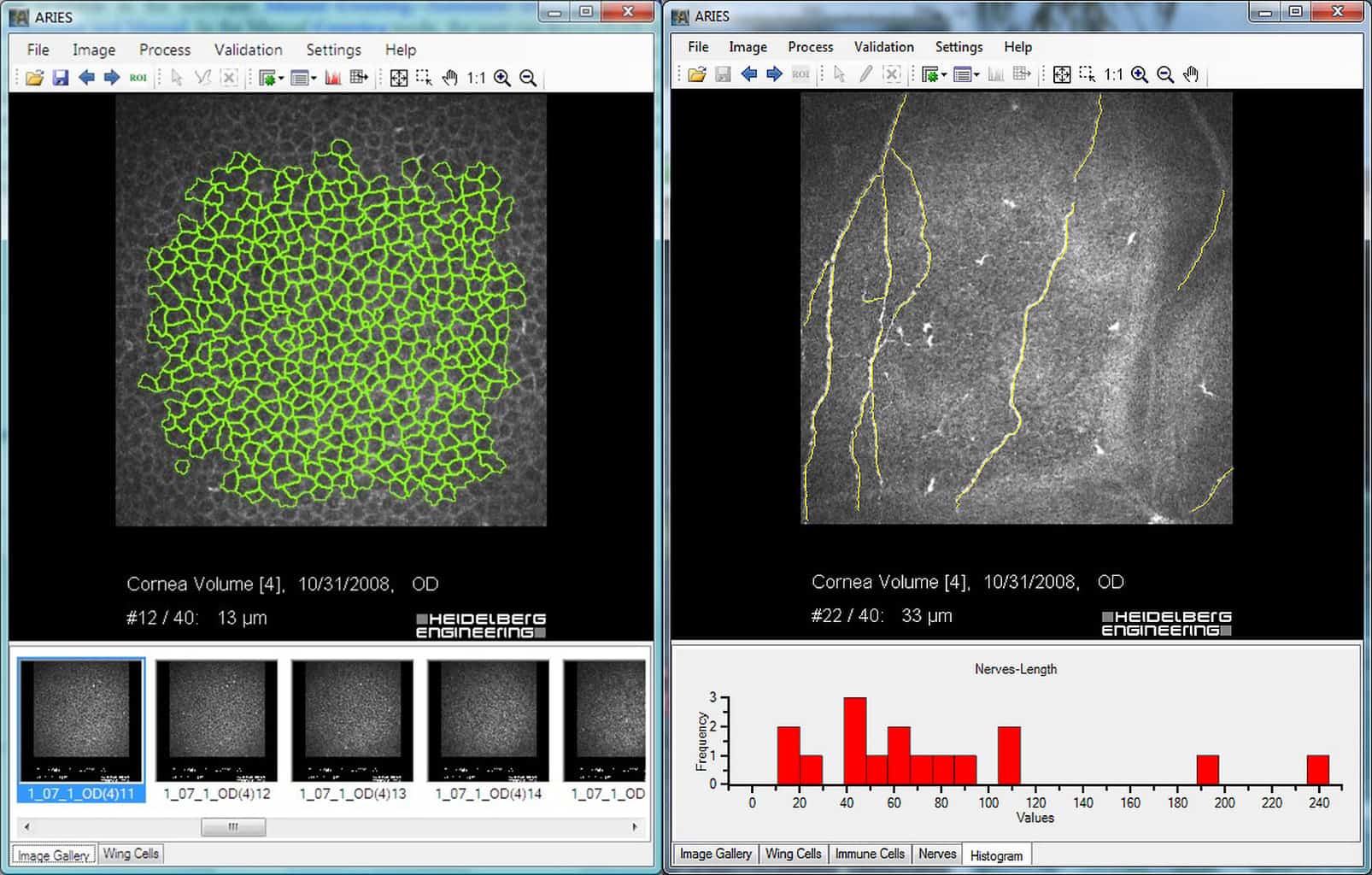
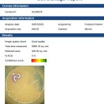
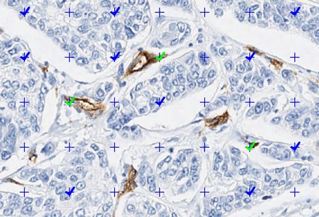
Aries
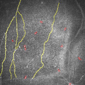
The epithelial layer of corneal cells is rich in clinical information. Aires automatizes the detection of nerves, neuromas, immune cells, and polyhedral cells in confocal imaging of the cornea
ADCIS solutions for analyzing images of the eye surface

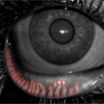
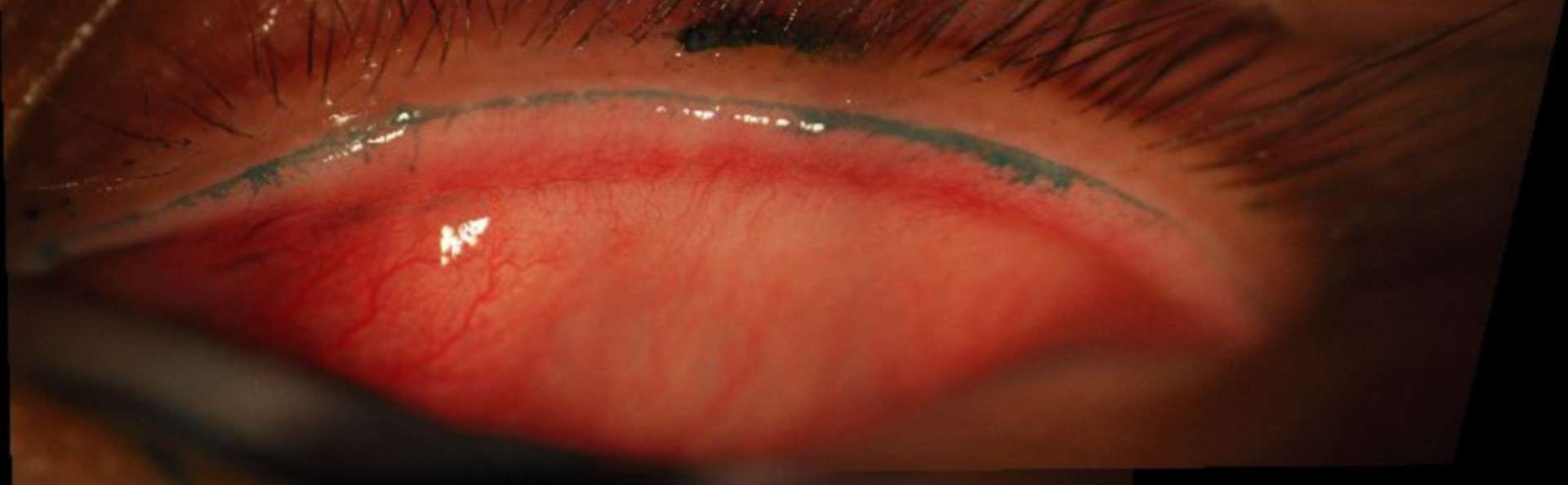
RedAI
Automatic vessel segmentation, quantification, and grading of eye redness
DryAI
Dry eye assessment varies too much between examiners. DryAI standardizes every measurement meibomian glands, tear meniscus, TBUT for reliable longitudinal tracking.
ADCIS solutions in contact lens fitting
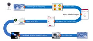
Contactology patient record
Scleral lenses prescription is a complex workflow. ADCIS Freeform Designer simplifies the prescription of custom scleral lenses.
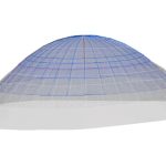
3D Lens viewer
A web platform for 3D display of custom lenses tailored to contact lens fitting
Custom Development | Have a specific imaging challenge? We build it
If an off-the-shelf solution doesn’t fit your workflow, ADCIS builds it from scratch.
From initial audit to production deployment, our team of engineers and PhDs handles the full development cycle combining computer vision, deep learning, and 30 years of ophthalmic domain expertise.
We’ve built 100+ custom applications for industry partners, medical device manufacturers, and pharma corporations.
We’ll build yours too.
Research | Where the next breakthroughs are built
ADCIS doesn’t just ship products, we push the science forward. Through collaborative research programs with leading academic and clinical institutions, we develop the AI algorithms that become tomorrow’s clinical tools.
Current flagship: EviRed a national program to reclassify diabetic retinopathy using AI and multimodal imaging. 3,000 patients. 12 centers. ADCIS at the core.
Since 2021, ADCIS has been contributing to the EviRed project, one of the most ambitious RHU projects in ophthalmology. EviRed uses artificial intelligence and multimodal imaging to establish a new classification of diabetic retinopathy in order to improve patient diagnosis, prediction, and treatment:
– Artificial intelligence: combines the deep learning expertise of the LATIM laboratory and ADCIS engineers
– Multimodality: includes ultra-wide field fundus photography, optical coherence tomography, and OCT angiography
– Recruitment of a cohort of 3,000 patients by 12 recruitment centers and follow-up of patients for 2 years
– A dedicated reading center has been created to support the project and annotate the images that will be used to train the deep learning algorithms
The roles of ADCIS and Evolucare in EviRed consist of providing the entire diagnostic process: annotation software, specialized multimodal viewers, deep learning algorithms, and the project’s IT infrastructure
Other innovative ADCIS projects in ophthalmology
Alongside its clinical, academic, and institutional partners, ADCIS continues to invest in innovation to improve the screening, monitoring, and prediction of the progression of ophthalmic diseases.
DiabEyeAI
Screening for diabetic retinopathy in pharmacies, as close as possible to the patient
SEDAAR
Multimodal screening for retinal diseases (OCT, retinography)
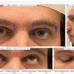
Blepharoplasty
Software for automatic detection of key points in the periorbital region to assess the impact of eyelid surgery
Messidor, the retinal image database dedicated to research
ADCIS hosts the image database from the Messidor project (Méthodes d’Évaluation de Systèmes de Segmentation et d’Indexation Dédiées à l’Ophtalmologie Rétinienne). Developed to support studies on computer-assisted diagnosis of diabetic retinopathy, the Messidor database consists of 1,200 color fundus images and is available to the public.

