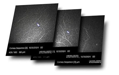
Confocal image processing of corneal cells layers
ARIES (AlConfocal Rapid Image Evaluation System) is a software developed by ADCIS to automatically process images of the cornea acquired by a confocal microscope. This powerful diagnosis and research application aims at automatically identifying, segmenting and quantifying nerves and cells of the corneal epithelium.
ARIES is widely used all over the word by research groups, CROs, pharmaceuticals and biotech companies.
Confocal microscopy allows for high resolution, reliable, real time, imaging of the living corneal microstructure including normal corneal morphology, pathogen invasion, and degenerations, post surgical management, dry eyes, drug toxicities, endothelial monitoring and contact lens related changes.
The software has been designed from the ground to address a typical analysis of the cornea using a confocal microscope. It analyses cellular structures in the images and generate accurate measurements that are used for medical purposes.
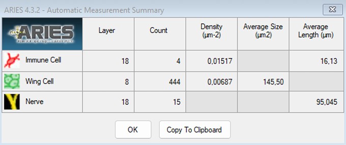
The software enables a quantified characterization of the corneal cells
Extraction of the image in which the nerves are the most pronounced
The software was designed to detect first, the section of a 3D volume that contains most of the nerve plexus, then to detect and analyze the cells present in the volume from the previously detected section. Nerves in the confocal images appeared as white objects (on a dark background) with an elongated and thinned shape. Therefore, image processing operators are designed to automatically detect the layer that contains such bright and thin
objects. The resultant identified layer is called the best nerve layer image (BNL).
After determination of the best nerve layer image, the software prompts the user to validate the section selection, and eventually to manually select another one after a visual inspection of neighbored sections.
Consequently, the newly selected image would be used to set the parameters for the detection of epithelial cells.
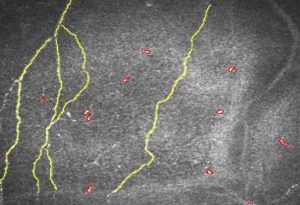
Automatic identification of the best nerve layer
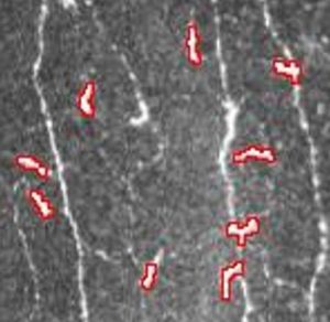
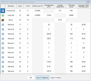
Analysis of Immune Cells
The immune cells were detected and analyzed in the best nerve layer image. Immune cells appeared in the images as small, white, and usually compact
objects. These cells could occur anywhere in the best nerve layer image: either isolated from the nerves or connected to the nerves (appearing as small branches on the nerve network). In order to analyze these immune cells, the nerves in the same image had to be excluded from the analysis.
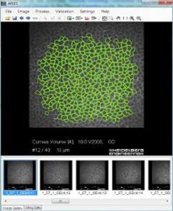
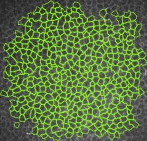
Analysis of Epithelial Cells
Epithelial cells (in the center of the epithelium) were targeted for analyses, rather than basal epithelial cells (most posterior in epithelium) or superficial epithelial cells (most anterior in epithelium), for a number of reasons.
Basal epithelial cells may present the following problems in analyses:
- Indistinct cell borders in confocal micrographs, with borders becoming more pronounced incorneas with edema.
- Cellular density that is relatively consistent among some types of corneal disorders, making this layer an unreliable marker of corneal health.
- Density that appears to vary between types of confocal microscopes and between studies.
For these reasons, epithelial cells were chosen for analysis: these cells are the most representative of corneal health because the measures have associated medical and automatic direction detection is relatively easy and very reliable
(thanks to the well-marked cell borders).
New ! ARIES neuroma module
Released in 2025, this new module enables an automatic and quantitative analysis of corneal neuromas
ARIES now integrates an advanced deep learning module for automatic detection, segmentation and quantification of corneal neuromas. Leveraging a clinically validated AI model trained on thousands of In Vitro Confocal Microscopy images, this module provides fast, quantified and reproducible measurements of neuroma morphological characteristics.
It enhances diagnostic confidence, supports longitudinal follow-up, and reduces manual analysis time from minutes to seconds, enabling clinicians to better evaluate neuropathic corneal pain, dry eye disease, diabeticneuropathies and post-surgical recovery.
Corneal microneuromas are powerful markers for various ocular or systemic diseases. Their automatic detection, segmentation, and quantification using the ARIES neuroma module facilitates a range of clinical analyses:
- Early and reproducible screening for neuropathic corneal pain
- Objective monitoring of postoperative neuropathic complications
- Detection of nerve damage caused by inflammation
- Systematic screening of the nerve network, useful for any differential diagnosis
Main benefits of the neuroma module
- Automatic and accurate detection and segmentation of neuromas using deep learning in a matter of seconds, natively integrated into ARIES
- Reproducibility and objectivity of results that cannot be achieved manually, for consistent results across an entire population
- A tool suitable for research, clinical studies, and screening programs
Downloads:
MORE INFORMATION
For any demonstration or explanation requests, please feel free to contact us via the button below.