
ARIES
From Corneal Confocal Images to Quantitative Biomarkers.
Automatically.
ARIES Platform for Corneal Confocal Microscopy Analysis
Transform confocal microscopy images into actionable clinical insights.
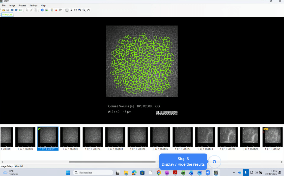
ARIES (AlConfocal Rapid Image Evaluation System) is an advanced software solution developed by ADCIS for the automated analysis of corneal confocal microscopy images. It enables fast, objective and reproducible quantification of corneal biomarkers for research and clinical studies.
ARIES automatically identifies, quantifies, segments and measures key corneal structures, enabling researchers and clinicians to obtain robust and reproducible results in seconds. Its automated workflow increases observations reproductibility while increasing productivity and confidence in image-based assessments.
Typical Biomarkers Analyzed
- Corneal nerve fiber density
- Corneal nerve branch density
- Immune cell metrics
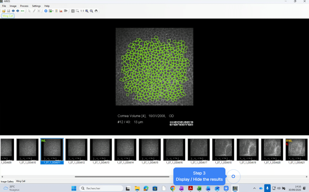
- Epithelial cell metrics
- Corneal neuroma metrics
Why Researchers Choose ARIES?
THOUSANDS
of clinically validated images
SECONDS
to complete an analysis
MEASURED
morphological metrics
REPRODUCIBLE
quantitative biomarkers
Key Features:
- Automated detection of corneal nerves, immune cells, epithelial cells, neuromas.
- Quantitative measurements and biomarker extraction.
- Manual review and correction tools when required.
- 2D and 3D image analysis capabilities.
- Exportable data for statistical and clinical research workflows.
Key Benefits:
- Analysis completed in seconds
- Trained on thousands of validated images
- Automated biomarker extraction
- Consistent measurements across operators
Scientifically Validated and Peer-Reviewed
ARIES has been developed in close collaboration with leading ophthalmology researchers and is supported by peer-reviewed scientific publications.
The platform provides standardized and reproducible measurements that help researchers generate reliable evidence from corneal confocal microscopy data.
- Peer-reviewed publication in Cornea
- Developed with leading ophthalmology researchers
- Thousands of annotated images used for AI training
- Reproducible and standardized measurements
Who uses ARIES?
- Contract Research Organizations (CROs): Accelerate image analysis and standardize biomarker generation across multicenter studies.
- Pharmaceutical companies: Generate reliable quantitative endpoints for clinical research and drug development.
- Research institutes: Automate image processing workflows while improving data consistency and reproducibility.
- Ophthalmology centers: Support advanced corneal imaging studies with AI-powered analysis tools.
NEW MODULE NEUROMA
Deep Learning-Based Corneal Neuroma Detection and Quantification
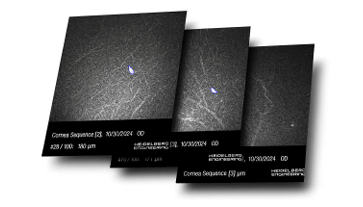
The ARIES Neuroma Module introduces an innovative deep learning-based approach for the automated detection, segmentation, and quantification of corneal neuromas.
Developed through the long-standing collaboration between ADCIS and the SERI (Singapore Eye Research Institute), this module was trained on thousands of clinically validated confocal microscopy images to deliver objective and reproducible neuroma measurements.
By transforming what was traditionally a complex and operator-dependent task into a rapid and standardized workflow, the Neuroma Module enables researchers and clinicians to unlock the full potential of neuroma-based biomarkers.
Applications
- Diabetic neuropathy research
- Drye eye disease studies
- Neurotrophic keratopathy assessment
- Clinical trials
- Drug development programs
- Ophthalmology research
Key Features
- Automated AI-based neuroma detection and quantification
- Analysis completed in seconds rather than minutes
- Reduced inter-observer variability
- Standardized biomarkers for clinical trials and research studies
- Seamlessly integrated within the ARIES platform