

AI-Powered Corneal Endothelium Analysis
for Objective and Reproducible Assessments
CornAI
The corneal endothelium plays a critical role in maintaining corneal transparency and visual function. Yet assessing endothelial cell health remains a complex and time-consuming task, often relying on manual or semi-automated measurements that are prone to variability.
CornAI leverages advanced image analysis and artificial intelligence to automatically analyze specular microscopy images and generate objective, reproducible, and clinically meaningful endothelial biomarkers.
By transforming corneal images into quantitative data, CornAI helps clinicians, researchers, eye banks, and industry partners improve diagnostic accuracy, standardize evaluations, and support data-driven decision-making.
Turn your endothelial images into actionable clinical insights in seconds, with consistent and reliable results.

From Subjective Evaluation to Quantitative Corneal Biomarkers
Reliable assessment of endothelial cell morphology and density is essential for patient follow-up, corneal disease management, transplantation, and clinical research.
Traditional approaches often present limitations:
- Manual counting is time-consuming
- Results may vary between operators
- Longitudinal comparisons can be difficult
- Large datasets require significant human effort
CornAI helps organizations:
- Automate endothelial cell analysis
- Improve measurement reproducibility
- Standardize corneal assessments
- Generate objective imaging biomarkers
- Support clinical decision-making
- Accelerate research studies
- Facilitate longitudinal patient monitoring
- Reduce manual analysis workload
With consistent quantitative measurements, CornAI increases confidence in both clinical and research outcomes.
Designed for Corneal Specialists, Researchers and Industry
CornAI combines artificial intelligence, image analysis, and ophthalmology expertise to simplify corneal endothelium evaluation.
Key Capabilities
- Automated endothelial cell detection
- Cell density quantification
- Polymegathism and pleomorphism analysis
- Morphological characterization
- Objective image quality assessment
- Longitudinal follow-up capabilities
- Standardized quantitative reporting
- High-throughput image processing
- AI-assisted decision support
- Exportable clinical and research metrics
The result is a reliable and scalable solution suitable for both routine clinical practice and advanced research applications.
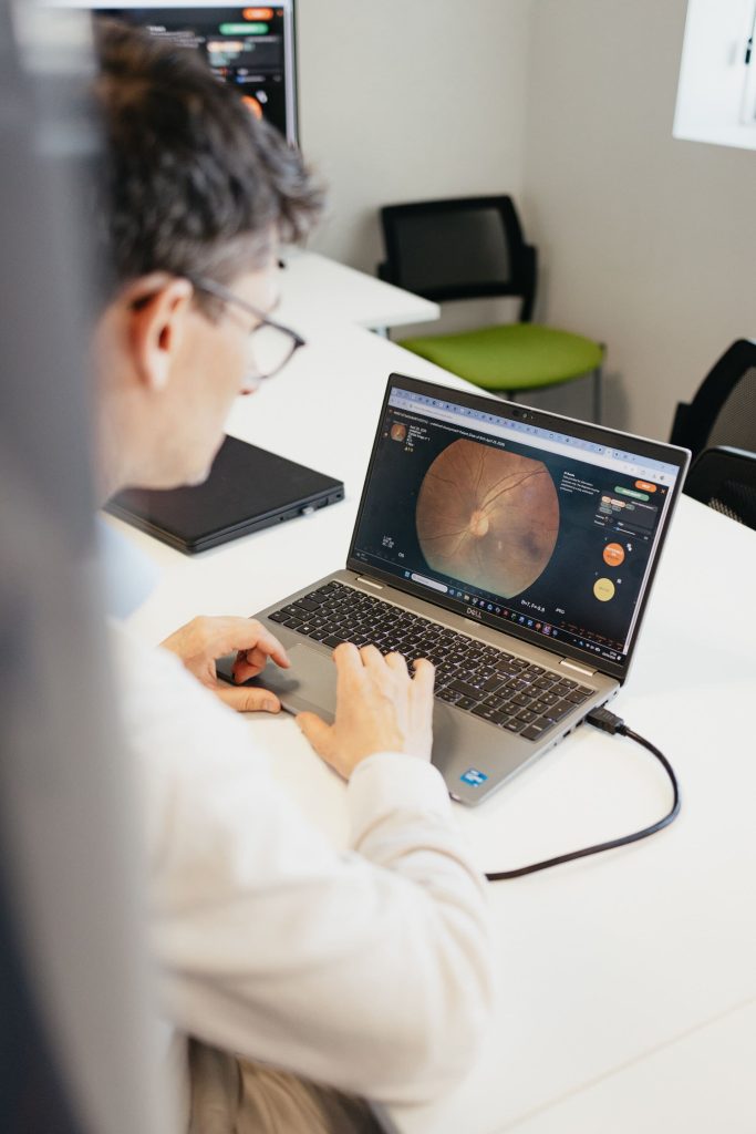
Who Benefits from CornAI ?
From Corneal Images
to Clinical Intelligence
CornAI enables ophthalmology professionals to exploit the full value of specular microscopy data. Whether monitoring endothelial health, evaluating corneal diseases, qualifying donor tissues, or generating endpoints for clinical trials, CornAI transforms image-based assessments into robust, quantifiable information.
From image acquisition to clinical interpretation, CornAI helps deliver more objective, reproducible, and actionable corneal evaluations.
- Fully automated endothelial analysis
- Objective and reproducible measurements
- AI-powered cell characterization
- Standardized quantitative biomarkers
- Designed for clinical and research environments
- Supports longitudinal patient monitoring
- Compatible with modern ophthalmic workflows
- Built on more than 30 years of ADCIS expertise in image analysis and computer vision
- Corneal Endothelium Assessment
- Endothelial Cell Density Analysis
- Fuchs Dystrophy Monitoring
- Post-Surgical Corneal Follow-Up
- Corneal Research Programs
- Eye Bank Tissue Evaluation
- Clinical Trial Imaging Endpoints
- AI-Assisted Ophthalmic Imaging
Scientifically Validated Performance
Developed in collaboration with leading international ophthalmology experts, CornAI has been scientifically validated through a peer-reviewed publication in the British Journal of Ophthalmology.
The solution automatically analyzes specular microscopy images and delivers standardized corneal endothelial measurements, even in the presence of complex images or pathologies such as Fuchs endothelial corneal dystrophy.
Key Benefits of CornAI
- Peer-reviewed scientific publication in the British Journal of Ophthalmology
“Development and validation of a deep learning model for automatic assessment of corneal endothelium” - Developed and validated using nearly 900 specular microscopy images from both healthy and pathological corneas, including independent external validation.
- Automated analysis of 8 key corneal endothelial parameters, including cell density, coefficient of variation, pachymetry, mean cell area, standard deviation, hexagonality, and minimum and maximum cell area.
- Robust performance across varying image quality levels and in challenging conditions such as Fuchs dystrophy with guttae.
- Results generated within seconds, with accuracy comparable to reference methods used in clinical practice.
CornAI transforms specular microscopy into reliable quantitative data for clinical research and ophthalmic practice.
See Beyond Cell Counts. Access Meaningful
Corneal Biomarkers.
Improve efficiency, standardization, and confidence in endothelial
assessments with AI-powered image analysis.